1 Introduction
More than 30% of all human proteins contain unfolded regions (). This stands in marked contrast to how little we know about them (). Intrinsically disordered proteins (IDPs) lack a stable tertiary structure, and thus lack a hydrophobic core. They thus also lack an active site. IDPs often interact with other proteins by means of short linear motifs (SLiMs), which are very abundant in disordered regions. This is a fundamental component of cell signalling (). These IDP characteristics have many consequences for aligning their sequences and thus for their study in general. Algorithms underlying existing MSA software are directly or indirectly based on knowledge obtained from studying 3D protein structures. Aligning a hydrophobic residue in the protein core with a hydrophilic one is penalized heavily when aligning ordered proteins, but this is much less the case when aligning IDPs simply because IDPs do not have a hydrophobic core. In addition, folded proteins contain large protein–protein interaction surfaces, ion binding sites, active sites or other 3D motifs that MSA software capitalizes on, while IDPs generally consist of short motifs surrounded by stretches of highly variable length and composition. The main function determinants of IDPs are SLiMs, and posttranslational modifications (PTMs).
For the specific purpose of aligning IDPs, we introduce Knowledge-based multiple sequence alignment (MSA) for IDPs, or KMAD, that incorporates SLiM, domain and PTM annotations in the alignment procedure. Obviously, the inclusion of this knowledge will cause these motifs to line up in the final MSA without certainty that this reflects biological reality. This way, however, KMAD generates hypotheses that can be validated experimentally to progress, for example, protein engineering, drug design or the analysis of genetic disorders. In these research fields, scientists most often are interested in one single protein and want to gather information for this one protein. In the particular case of IDPs such information normally relates to SLiMS and PTMs and how conserved these are in an MSA. In the HSSP project (), the concept of the insertion-free MSA was introduced specifically for this purpose (see Fig. 1). Although KMAD can produce ‘normal’ MSAs (i.e. with insertions and deletions possible in all sequences), we tend to use it insertion-free when studying IDP MSAs.
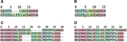
Fig. 1
Hypothetical example of an insertion free alignment. (A) ‘normal’ MSA. (B) The same alignment, but with the insertions removed from the first sequence. Obviously the residues in the other sequence that aligned with the removed insertions were removed too. The residues g and l are in lower case characters to indicate that an insertion was removed between them. (C, D) Illustrating how the removal of insertions from the sequence of interest can increase the amount of information that can be extracted from the MSA. In (C), the SVIDL motif is duplicated in the second sequence. In the third and fourth sequences these motifs are more similar to the second instance of the motif in sequence two than to the first instance. By removing the insertion we learn that the SVIDL motif in the first sequence indeed is conserved, and any knowledge available for these residues in the bottom two sequences can be transferred to the first sequence
MSA validation is a hotly debated issue that seems mostly unsolved (). Even worse, for the same set of sequences different MSAs can be produced that all seem correct (). Alignments extracted from structure superpositions are generally considered the gold standard, but many examples exist in which a structure superposition-derived MSA does not reflect well the sequence family’s phylogeny (). The MSA benchmark suites BAliBASE (; ) and PREFAB () are mostly structure superposition based, albeit that superpositions used in these suites do not always agree () with external sources such as CATH () or SCOP (). It is not clear how these discrepancies arose but it is known that the same structures sometimes can be superposed differently depending on the algorithm used and the parameter choices (; ). recently studied quality measures for protein alignment benchmarks and concluded that ‘protein alignment assessment is more challenging than generally realized’; he also concluded that BAliBASE block identifications do not correlate well with conserved protein secondary structures. We used BAliBASE and PREFAB to validate KMAD’s MSAs and the MSAs produced by a series of well-known alignment programs, and conclude that the differences in alignment validation score between the six methods are smaller than the ‘noise’ in BAliBASE (www.cmbi.ru.nl/kmad/balibase/). It should be noted that KMAD was designed to produce insertion-free alignments (see Fig. 1) that best allow for transfer of information from the whole alignment to the one sequence of interest while BAliBASE and PREFAB contain ‘complete’ alignments that better reflect the underlying phylogeny.
2 Methods
E
The KMAD server can annotate IDPs in the MSA. IDPs are either obtained from the D2P2 database of disorder predictions (), or by running the freely available disorder predictors: GlobPlot (), DISOPRED (), SPINE-D (), PSIPRED (), PreDisorder () and IUPred ().
KMAD was designed for the optimal alignment of IDPs. Proteins with a stable tertiary structure (non-IDPs) often also contain PTMs, SLiMs and domains. KMAD can be used for the alignment of non-IDPs too, but better results will be obtained if non-IDPs are first aligned with software optimized for that task (i.e. any software other than KMAD) followed by fine-tuning the resulting alignment with KMAD. We call this the refinement option of KMAD.
MSA quality was evaluated in two ways. First, MSAs made with Clustal Omega and KMAD were visually inspected in light of the known biology. Second, a quantitative analysis was performed with the protein linear motif benchmark for MSA tools from the BAliBASE suite, and with the PREFAB suite.
3 Implementation
i
jprofileimiDiDj.fti.iRfijj
RfRf
4 Results
KMAD overcomes several problems encountered with other methods when aligning IDPs. Two examples are shown in Figures 3 and 4. The first example (Fig. 3) illustrates the advantage of KMAD’s use of SLiMs. Clustal Omega spreads out the LIG_BIR_II_1 motif. In the KMAD alignment, the motif is lined up nicely in all sequences but 2. Even if this predicted SLiM should not be aligned as in the B panel of Figure 3, the bioscientist will still benefit from awareness of the presence of this conserved motif in the N-terminal segment.
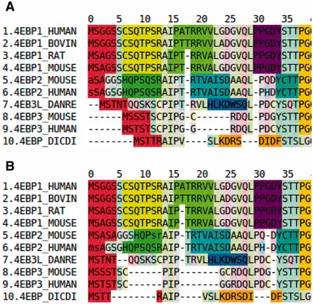
Fig. 3
Excerpts from Clustal Omega and KMAD alignments of eukaryotic translation initiation factor 4E-binding protein 1. (A) Clustal Omega alignment annotated with motifs; (B) KMAD alignment annotated with motifs. Each of the bright colours represents a different motif (a bright red background indicates the LIG_BIR_II_1 motif; further colour details are given at the project website)
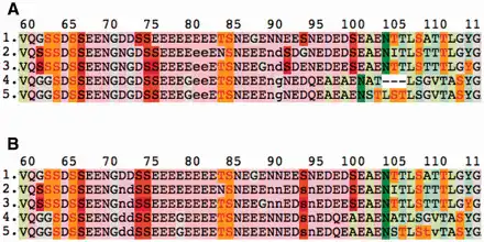
Fig. 4
Excerpts from Clustal Omega and KMAD alignments of human sialoprotein (SIAL_HUMAN) with four homologues. (A) Clustal Omega alignment annotated with PTMs; (B) KMAD alignment annotated with PTMs. PTM sites are highlighted with bright colours (red is annotated phosphorylation, orange is predicted phosphorylation, green is N-linked glycosylation; further colour details are given on the project website). The two serines that seem to appear in the sequences 4 and 5 around position 93 in the KMAD alignment are located in the gap (indicated with lowercase characters) around position 90 in the Clustal Omega alignment. In DNA analysis, protein engineering, etc, this software feature is used frequently. The gapped alignment that provides a more phylogeny-oriented view on this sequence family is available at the KMAD website
The second example (Fig. 4) illustrates the use of PTMs. The phosphorylated serines at positions 74 and 75 in the first sequence are shifted to the left relative to the columns of phosphorylated serines at positions 75 and 76 in the other four sequences. A gap at position 73 in the query sequence would be a better solution in this case, but because we want to keep the first sequence indel-free, an insertion is introduced in all other sequences. The phosphorylated serine at position 94 in the first sequence seems better aligned with the serines at position 92 in sequences 2 and 3, and indeed, KMAD finds this solution.
Eventually, glycosylated asparagines at position 102 in sequences 4 and 5 should most probably be aligned to asparagines at position 105 from sequences 1–3. All problems mentioned above are solved by KMAD with no harm to the rest of the alignment. The problem that the predicted threonine phosphorylation sites around position 106 cannot be lined up is beyond KMAD’s reach.
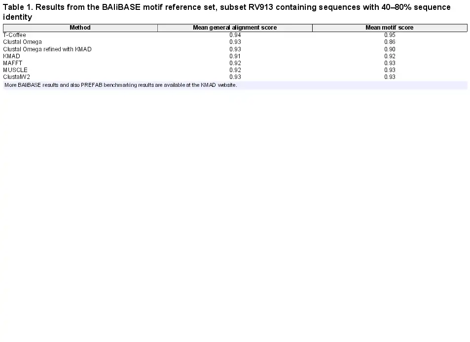
Figures 3 and 4 provide two examples in which knowledge of the presence of sequence motifs is used to better align those motifs. This method is of course highly cyclic and we therefore compared the performance of KMAD with five other MSA methods using the BAliBASE and PREFAB suites. Most proteins in these benchmarks have a stable 3D structure, while KMAD was designed for the alignment of IDPs. We therefore used KMAD in refinement mode starting with Clustal Omega alignments. To avoid circularity when testing KMAD on the BAliBASE motif reference set we performed a leave-one-out validation, i.e. in each alignment the motif annotated by BAliBASE was excluded from the KMAD annotations.
The score differences in Table 1 are all within the margin of error, given that noise in the benchmarks (www.cmbi.ru.nl/kmad/balibase/) contributes a few percent to the scores.
We chose four sequence families for testing the CPU performance of the six methods. The sequences in these families on average are about 150, 250, 550 and 750 amino acids long, respectively. From each family subsets of 5, 50, 500, 5 K, 20 K and 80 K were selected and these 24 groups of sequences were aligned with each of the six methods using each time one core on the same computer that had more than adequate RAM, so that the wall-time of each calculation provided a good measure for the CPU performance of the method. The results are visualized in Figure 5. In this plot, some points are missing because we stopped each alignment when after 1 week no result had been returned yet. Clearly, KMAD outperforms the other methods for large alignments.
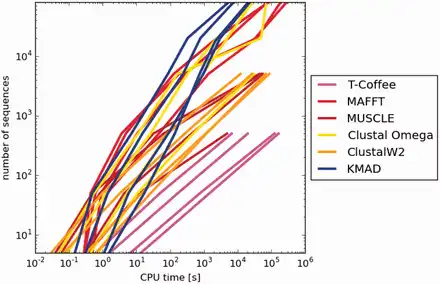
Fig. 5
Log–log plot of CPU performance. The abscissa and ordinate are logarithmic scales for the CPU time and the number of sequences, respectively. For every method there are four curves each representing a different protein family
5 Discussion
In the field of sequence alignment research it is common practice to compare the sequence alignments obtained with MSA software with those that are obtained from structure superpositions (). IDPs do not possess a static 3D structure so that this method is not applicable here. We have discussed several examples in which KMAD produces IDP alignments that intuitively feel correct, and several more examples are worked out in detail at the associated website. It should be noted, however, that the features used for alignment quality determination are the same as those used for producing the MSA. This is not a very elegant method, but given the nature of IDPs probably the best that can be done. KMAD certainly will emphasize functionally important IDP residues and regions, and thus will provide a basis for subsequent experiments needed to shed light on the sequence structure function relation of this intriguing branch of the protein kingdom.
6 Usage
KMAD is available as a standalone version and as a web-server. The standalone version consists of three parts to (1) obtain information about the features (IDPs, PTMs, SLiMs and domains) and map them onto the sequences; (2) KMAD itself to use all information to align the sequences; and (3) the output scripts. These programs are available with installer, documentation, etc., from the projects website. KMAD accepts a query sequence and runs its whole pipeline automatically to predict disordered regions, align sequences or annotate alignments. The standalone version and the web-server both allow user to change parameters, including user-defined features. A REST API is available, so that users can access KMAD programmatically without the need to install the standalone version.
Acknowledgements
We thank the NewProt project that is funded by the European Commission within its FP7 Programme, under the thematic area KBBE-2011-5 with contract number 289350. Toby Gibson, Des Higgins and David Jones carefully read the manuscript and provided advice.
References
- Altschul S., et al.. (1990) Basic local alignment search tool. J. Mol. Biol., 215, 403–410.
- Andreeva A., et al.. (2008) Data growth and its impact on the SCOP database: new developments. Nucleic Acids Res., 36(Suppl 1), D419–D425.
- Ashburner M., et al.. (2000) Gene Ontology: tool for the unification of biology. Nat. Genet., 25, 25–29.
- Bateman A., et al.. (2004) The Pfam protein families database. Nucleic Acids Res., 32(Suppl 1), D138–D141.
- Blom N., et al.. (1999) Sequence and structure-based prediction of eukaryotic protein phosphorylation sites. J. Mol. Biol., 294, 1351–1362.
- Cheng J., et al.. (2005) Accurate prediction of protein disordered regions by mining protein structure data. Data Min. Knowl. Disc., 11, 213–222.
- Dinkel H., et al.. (2013) The eukaryotic linear motif resource ELM: 10 years and counting. Nucleic Acids Res., 42(Database issue), D259–D266.
- Dosztányi Z., et al.. (2005) IUPred: web server for the prediction of intrinsically unstructured regions of proteins based on estimated energy content. Bioinformatics, 21, 3433–3434.
- Edgar R.C. (2004) MUSCLE: multiple sequence alignment with high accuracy and high throughput. Nucleic Acids Res., 32, 1792–1797.
- Edgar R.C. (2010) Quality measures for protein alignment benchmarks. Nucleic Acids Res., 38, 2145–2153.
- Gibson T.J. (2009) Cell regulation: determined to signal discrete cooperation. Trends Biochem. Sci., 34, 471–482.
- Gotoh O. (1982) An improved algorithm for matching biological sequences. J. Mol. Biol., 162, 705–708.
- Iantorno S., et al.. (2014) Who watches the watchmen? An appraisal of benchmarks for multiple sequence alignment. Multiple Seq. Align. Methods, 1079, 59–73.
- Irving J.A., et al.. (2001) Protein structural alignments and functional genomics. Proteins, 42, 378–382.
- Jones D.T. (1999) Protein secondary structure prediction based on position-specific scoring matrices. J. Mol. Biol., 292, 195–202.
- Joosten H.-J. (2007) 3DM: from data to medicine. PhD Thesis, Wageningen University.
- Katoh K., et al.. (2002) MAFFT: a novel method for rapid multiple sequence alignment based on fast Fourier transform. Nucleic Acids Res., 30, 3059–3066.
- Konagurthu A.S., et al.. (2006) MUSTANG: a multiple structural alignment algorithm. Proteins, 64, 559–574.
- Larkin M.A., et al.. (2007) Clustal W and Clustal X version 2.0. Bioinformatics, 23, 2947–2948.
- Linding R., et al.. (2003) GlobPlot: exploring protein sequences for globularity and disordery. Nucleic Acids Res., 31, 3701–3708.
- Midic U., et al.. (2009) Protein sequence alignment and structural disorder: a substitution matrix for an extended alphabet. In Proceedings of the KDD-09 Workshop on Statistical and Relational Learning in Bioinformatics, ACM, pp. 27–31.
- Needleman S.B., Wunsch C.D. (1970) A general method applicable to the search for similarities in the amino acid sequence of two proteins. J. Mol. Biol., 48, 443–453.
- Nguyen K., Pan Y. (2013) A knowledge-based multiple-sequence alignment algorithm. IEEE/ACM Trans. Comput. Biol. Bioinform., 10, 884–896.
- Notredame C., et al.. (2000) T-Coffee: a novel method for fast and accurate multiple sequence alignment. J. Mol. Biol., 302, 205–217.
- Oates M.E., et al.. (2013) D2P2: database of disordered protein predictions. Nucleic Acids Res., 41(D1), D508–D516.
- Pentony M.M., Jones D.T. (2010) Modularity of intrinsic disorder in the human proteome. Proteins, 78, 212–221.
- Perrodou E., et al.. (2008) A new protein linear motif benchmark for multiple sequence alignment software. BMC Bioinformatics, 9, 213.
- Sander C., Schneider R. (1991) Database of homology-derived protein structures and the structural meaning of sequence alignment. Proteins, 9, 56–68.
- Sickmeier M., et al.. (2007) DisProt: the database of disordered proteins. Nucleic Acids Res., 35(Database issue), D786–D793.
- Sievers F., et al.. (2011) Fast, scalable generation of high-quality protein multiple sequence alignments using Clustal Omega. Mol. Syst. Biol., 7, 539.
- Sillitoe I., et al.. (2015) CATH: comprehensive structural and functional annotations for genome sequences. Nucleic Acids Res., 43(D1), D376–D381.
- The Uniprot Consortium (2014) Activities at the universal protein resource (UniProt). Nucleic Acids Res., 42(Database issue), D191–D198.
- Thompson J.D., et al.. (2005) BAliBASE 3.0: latest developments of the multiple sequence alignment benchmark. Proteins, 61, 127–136.
- Van der Lee R., et al.. (2014) Classification of intrinsically disordered regions and proteins. Chem. Rev., 114, 6589–6631.
- Venselaar H., et al.. (2010) Protein structure analysis of mutations causing inheritable diseases. An e-Science approach with life scientist friendly interfaces. BMC Bioinformatics, 11, 548.
- Vriend G. (1990) WHAT IF: a molecular modeling and drug design program. J. Mol. Graph., 8, 52–56.
- Ward J.J., et al.. (2004) Prediction and functional analysis of native disorder in proteins from the three kingdoms of life. J. Mol. Biol., 337, 635–645.
- Zhang T., et al.. (2012) SPINE-D: accurate prediction of short and long disordered regions by a single neural-network based method. J. Biomol. Struct. Dyn., 29, 799–813.