SymbolMorphology research of the synovial membrane of the knee joint and clinical implications following synovial lesions
- Yong, Liu-jun
- Zhang, Xing-yu
1Department of Anatomy, Chengdu Medical College, Chengdu 610083, Sichuan Province, China; 2Troop 77156 Hospital of Chinese PLA, Leshan 614000, Sichuan Province, China
Yong Liu-jun, Lecturer, Department of Anatomy, Chengdu Medical College, Chengdu 610083, Sichuan Province, China [email protected]
Supported by: a grant from Sichuan Education Department, No. 08zc040*
Received: 2009-02-20
Accepted: 2009-07-20

Symbol
No caption available.

Symbol
No caption available.
Symbol
No caption available.
Symbol
No caption available.

Symbol
No caption available.
Symbol
No caption available.

Symbol
No caption available.
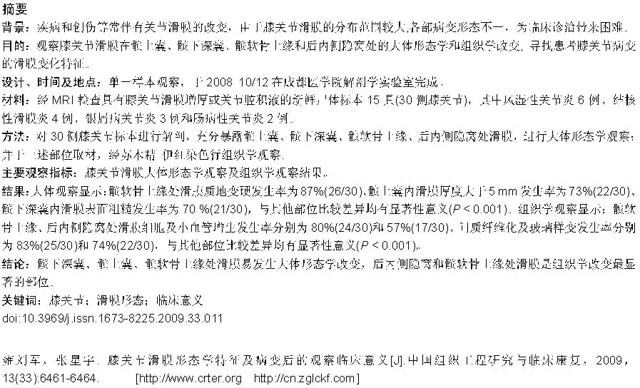
Abstract
BACKGROUND:
The majority of joint diseases and injuries are often accompanied with synovial changes. As the synovial membrane is distributed widely within the knee joint, pathological changes at different sites are different, leading to the difficulty in clinical diagnosis and treatment.
OBJECTIVE:
To observe the general morphologic and histological changes of the synovial membrane of the knee joint at the suprapatellar bursa, deep infrapatellar bursa, superior border of patella-cartilage and post-wall crypt, and to explore the synovial changing characteristics following patients' knee joint lesions.
DESIGN, TIME AND SETTING:
The observation of a single sample was performed in Department of Anatomy, Chengdu Medical College from October to December 2008.
MATERIALS:
Fifteen fresh cadaver specimens with the knee joint synovial thickening or joint effusion confirmed by MRI were selected, of which 6 cases were rheumatoid arthritis, 4 cases were tuberculous synovitis, 3 cases were psoriatic arthritis and 2 cases were enteropathy arthritis.
METHODS:
Bilateral knee joints (30 sides) of 15 cadaver specimens were anatomized to expose the suprapatellar bursa, deep infrapatellar bursa, superior border of patella-cartilage and post-wall crypt for general morphologic observation, and then the above specimens were observed histologically after hematoxylin-eosin staining.
MAIN OUTCOME MEASURES:
The results of general morphology and histology observation.
RESULTS:
The general observation revealed that 26 of the 30 sides were present with synovial hardening at the superior border of patella-cartilage, accounting for 86.67%, 22 of the 30 sides (73.33%) present with synovial thickness > 5 mm at the suprapatellar bursal, and 21 of the 30 sides (70 %) present with synovial roughness at the deep infrapatellar bursal. Every index of the rest parts had a significant deviation (P < 0.001). Histology observation showed that, at the superior border of patella-cartilage and post-wall crypt, hyperplasia incidences of synovial cells and small vessels were 80 %(24/30)and 56.67%(17/30), respectively, and interstitial fibrosis and hyalinization was 83.33% (25/30) and 73.53%(22/30), respectively, which significantly differed from other parts(P < 0.001).
CONCLUSION:
Synovial membrane at the deep infrapatellar bursa, suprapatellar bursa, and superior border of patella-cartilage was easily changed in morphology, and the histological variation was often found in the superior border of patella-cartilage and post-wall crypt.
Yong LJ, Zhang XY. Morphology research of the synovial membrane of the knee joint and clinical implications following synovial lesions.Zhongguo Zuzhi Gongcheng Yanjiu yu Linchuang Kangfu. 2009;13(33): 6461-6464. [http://http://www.crter.cn http://en.zglckf.com]
Symbol
No caption available.