Introduction
A fibroepithelial polyp (FEP) is a benign lesion of mesodermal origin, which commonly occurs on the skin and less frequently in the genitourinary tract (most often in the proximal ureter). Fibroepithelial polyps of the head and neck region are very rare. To our knowledge, the literature includes only 2 reported cases of FEP arising independently in the external auditory canal (EAC), which involved female patients of 16 and 28 years of age., Here, we present a unique case of FEP found in the EAC of a 2-year-old child, and we discuss this pathology.
Case Report
A 2-year-old girl, with no history of prenatal or perinatal risks, was referred to a tertiary referral hospital due to a tumor in the left EAC. The polyp was found by the child’s parent after a regular bath. The girl was otherwise healthy, with no monitored disease or history of trauma, ear pain, secretion, or bleeding. No signs of hearing loss were observed, and newborn hearing screening using otoacoustic emission was negative (without suspicion of hearing loss).
Clinical examination revealed a smooth, slightly translucent, oval-spherical tumor with a diameter of 1 cm, filling the left EAC. The origin was not apparent (Figure 1). Findings on the right side were physiological. Computed tomography revealed no middle ear pathology and confirmed that the tumor was limited to the cartilaginous part of the left EAC without imminent cartilage erosion or spread to surrounding tissue. Mild peripheral contrast enhancement was observed. The tumor generally exhibited signs of benign behavior (Figure 2). The patient was indicated for surgical intervention under general anesthesia.
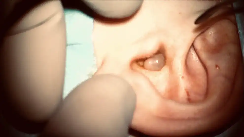
Figure 1
Otomicroscopy showing preoperative finding of a smooth, slightly translucent, oval-spherical tumor with a diameter of 1 cm, filling the left external auditory canal.
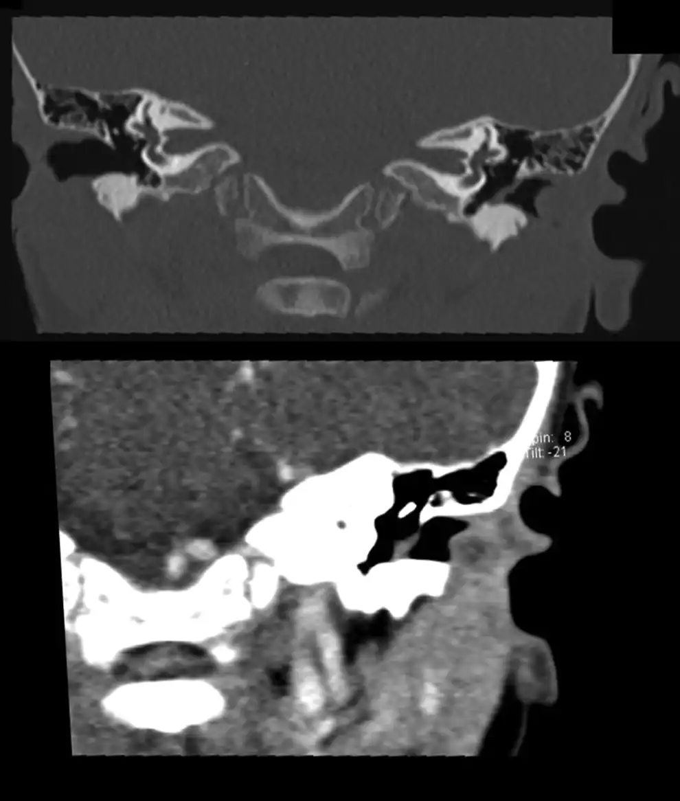
Figure 2
Preoperative computed tomography frontal scans before (upper image) and after (lower image) contrast medium application, revealing an oval tumor limited to the cartilaginous part of the left external auditory canal. These images show no imminent erosion of cartilage or spread to the surrounding tissue and mild peripheral contrast enhancement.
An endaural microscopic procedure was performed. The tumor was found to originate from the inferior wall of the cartilaginous part of the EAC. Complete resection was performed without complications. A substantial amount of detritus was sucked out from behind the tumor. The tympanic membrane was intact. Ear packing was inserted and then removed 1 day after surgery, after which the patient was discharged. Local antibiotic therapy was indicated for 7 days. The wound healed per primam. Histological examination confirmed the diagnosis of FEP (Figure 3). At 12 months postoperatively, the patient remains without symptoms and local clinical findings appear physiological.
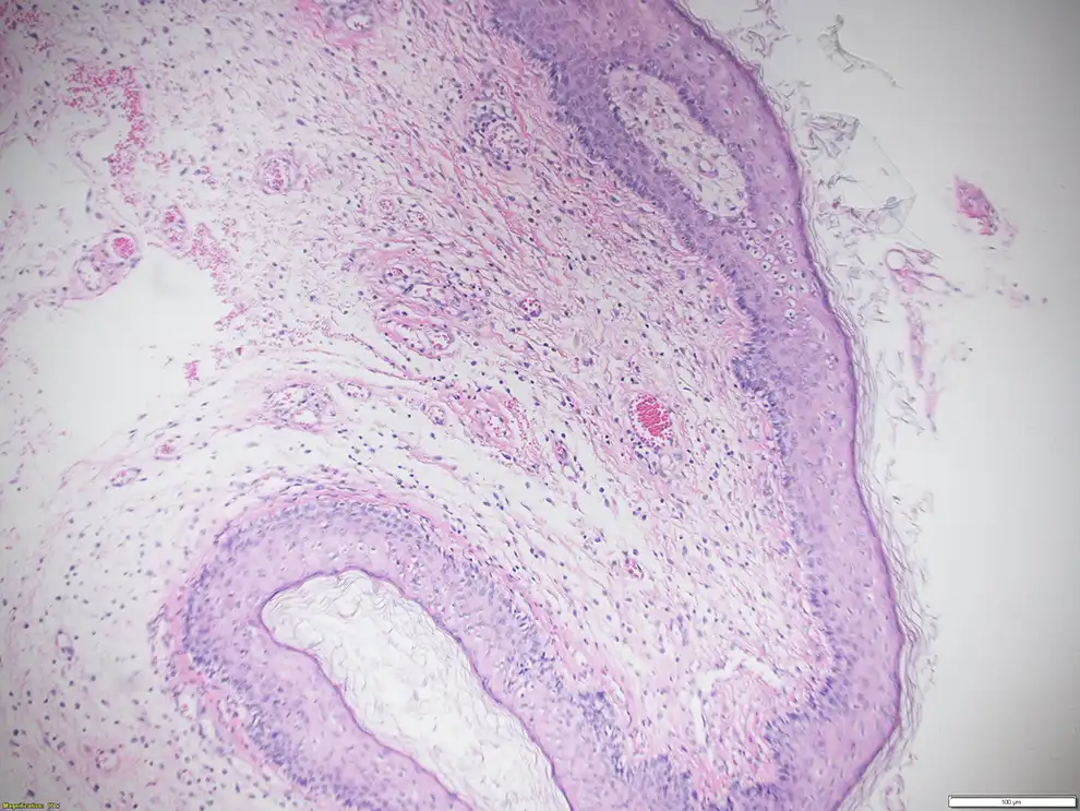
Figure 3
Histopathological findings of the fibroepithelial polyp with hematoxylin–eosin staining (×100 magnification).
Discussion
Fibroepithelial polyps are regarded as pseudotumors, and the pathophysiologic mechanism behind their development remains unclear. Their etiology is likely multifactorial, potentially involving chronic inflammatory process, chronic irritation from trauma or infection, carcinogens, hormonal imbalances, and congenital factors. The etiology of FEP in our case is unknown. The most probable options seem to be congenital factors or chronic irritation during ear cleaning by the parents. However, it is questionable whether sporadic irritation over a maximum duration of 2 years would be sufficient to cause the pathology. Additionally, a hormonal imbalance cannot be excluded, considering that all FEPs described in the EAC, to date, have been in females.
Fibroepithelial polyps are generally asymptomatic. However, problems may arise depending on the location. The presently reported patient would likely have soon experienced problems associated with EAC obstruction, considering the amount of detritus found behind the tumor at the time of surgery.
Tumor-like lesions of the EAC, the so-called “aural polyps,” include but are not limited to exostosis, osteoma, fibrous dysplasia, granuloma, ceruminous gland tumor, cholesteatoma, mycobacterial infection, papilloma, retained tympanostomy tubes, Langerhans cell histiocytosis, and malignancies. Final diagnosis is possible only after histopathological evaluation. Fibroepithelial polyp comprises a fibrovascular core surrounded by stratified squamous epithelium. Mast cells are frequently seen and likely interact with stromal cells to induce vascular and cellular differentiation. Immunohistochemical staining typically reveals strong desmin and vimentin positivity.
In our case, similar to the 2 previously reported cases, excision was the treatment of choice and provided a cure., Fibroepithelial polyp recurrence after resection is extremely rare in all regions. It is unclear whether it is crucial to have histology results before performing surgery, considering the wide spectrum of differential diagnoses of EAC tumor/polyp. In the previously reported cases, the authors knew the diagnosis before surgery. Although this is clearly beneficial, it must be noted that in those cases, verification was possible under local anesthesia since the patients were an adult and a teenager. Additionally, there is always a risk of excision of an inadequate or inconclusive sample. In our case, complete excision was indicated based on the preoperative findings from computed tomography scans, which excluded middle ear involvement or extracanal spread.
Conclusion
Here, we report a unique case in which an FEP was found in the cartilaginous part of the EAC of a 2-year-old child. The polyp was successfully treated by excision using an endaural approach and healed without complications. This is the very first report of an FEP in the EAC in the pediatric population. Although FEP is an extremely rare diagnosis, it should be considered in the differential diagnosis of a child’s EAC polyp.
Declaration of Conflicting Interests The author(s) declared no potential conflicts of interest with respect to the research, authorship, and/or publication of this article.
Funding The author(s) disclosed receipt of the following financial support for the research, authorship, and/or publication of this article: This work was supported by the Ministry of Health, Czech Republic—conceptual development of research organization (FNOs/2019).
Martin Formánek
https://orcid.org/0000-0002-5759-2073
References
- 1. Graff J, Patnaik S, Cohen T, Memo M. Ureteral polyp managed by endoscopic techniques. Rev Urol. 2019;21(1):45–48.
- 2. Tanaka N, Matsunobu T, Shiotani A. Fibroepithelial polyp of the external auditory canal: a case report and a literature review. Case Rep Otolaryngol. 2013;2013:818197. doi:10.1155/2013/818197
- 3. Thomas P, Rai P, Meena R. Fibroepithelial polyp of external auditory canal. Eur Ann Otorhinolaryngol Head Neck Dis. 2017;134(2):141–142.
- 4. Gliklich RE, Cunningham MJ, Eavey RD. The cause of aural polyps in children. Arch Otolaryngol Head Neck Surg. 1993;119(6):669–671.
- 5. Halvorsen TB, Johannesen E. Fibroepithelial polyps of the vagina: are they old granulation tissue polyps? J Clin Pathol. 1992;45(3):235–240.