Multinodular keratoacanthoma
a rare but definite entity
- Sardana, Kabir MD
- Sarkar, Rashmi MD
- Garg, Vijay Kumar MD
- Koranne, Ravindra V. MD
- Sharma, Ravi C. MD
- Sethi, Seema MD
A 62-year-old male farmer was seen at the skin outpatient department of the Lady Hardinge Medical College Hospital for evaluation of a large asymptomatic tumor on the right lower limb. It had been present for the last 11 months. The tumor began as a 2 × 1-cm sized lesion and progressively increased in size with central clearing and the appearance of multiple raised lesions on the periphery of the tumor. There was no history of trauma/infection at the site, exposure to tar/chemicals or excessive exposure to sunlight. There was no history of tuberculosis in the patient or family.
Dermatological examination revealed a hypertrophic, verrucous plaque of size 9 × 5 cm on the right shin with multiple nodules of size 1.5 cm to 2 cm on the periphery of the plaque with central clearing and scarring (Fig. 1). Regional lymph nodes were not enlarged, and remainder of the cutaneous examination was normal. A diagnosis of multinodular keratoacanthoma was made. A wedge biopsy was performed from the periphery of the plaque, and the histopathology confirmed the diagnosis of keratoacanthoma (Fig. 2).
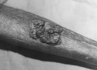
Figure 1
Multinodular hypertrophic plaque of size 9 × 5 cm on the right shin, with central clearing
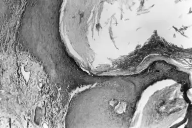
Figure 2
Marked hyperkeratosis with a central crater filled with keratin. The underlying epidermis shows acanthosis and hypergranulosis, while the papillary dermis reveals mild edema-congested blood vessels and chronic inflammatory infiltrate (H&E ×10)
Wide (4 mm) excisional surgery was performed and the defect was repaired with a full-thickness skin graft (Fig. 3). The patient was subsequently discharged and has been on regular follow up for the last 3 years with no recurrence.
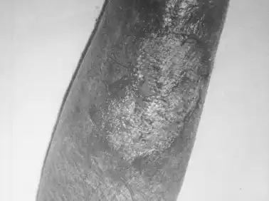
Figure 3
Full thickness graft in place following excisional surgery of the tumor