COVID-19 disease is an infection caused by the SARS-CoV2 virus. This can express a wide variety of different presentations, with up to 39% of the patients having ophthalmic manifestations.[] Follicular conjunctivitis has been reported by Sindhuja et al.[] in up to 8.66%, and eyelid manifestations have been reported by Meduri et al.[] in up to 38% of the patients. Many other ocular manifestations have been reported, including posterior segment involvement, but there is no publication reporting the lack of corneal epithelization after a corneal transplant.[]
A male infant with congenital glaucoma (known homozygous CYP1B1 mutation) status post Ahmed Valve OU underwent penetrating keratoplasty (PKP) for corneal opacification of the left eye at the age of 8 months. The patient tested negative for COVID-19 infection 2 days prior to surgery. The PKP was performed using the graft-over-host technique[] under general anesthesia with no complications. To trephine the recipient cornea, a 7.00-mm vacuum trephine was used and a 7.5-mm donor corneal graft was implanted. A total of 16 interrupted 10-0 Nylon sutures were placed to suture the graft in place, and antibiotic ointment and a pressure patch in the operated eye were placed at the end of the procedure. He was started on moxifloxacin drops (Vigamox®) every 4 h, prednisolone acetate drops (Pred Forte®) every 4 h, and cyclosporine 0.05% drops (Restasis®) 4 times a day. On postoperative day 1, the graft was clear with no signs of rejection or infection and the eye was quiet. On postoperative day 6, the child presented with bilateral eye swelling, runny nose, cough, and vomiting. Exam revealed severe bilateral conjunctivitis; however, the PKP was clear. The patient was evaluated in the emergency room, where he tested positive for the COVID-19 infection. The patient was admitted for 1 day for vomiting and respiratory symptoms. After discharge, he was frequently evaluated and the graft remained clear with no signs of infection or rejection.
The first exam under sedation (EUS) was performed 1 month after surgery (prior EUS could not be performed due to his COVID-19 infection). The right eye presented diffuse punctate epithelial erosions. The left eye revealed a lack of epithelialization in the graft [Fig. 1], with no signs of infection or rejection. The IOP was within the normal range. A dehydrated human amniotic membrane (BioDOptix®) was placed with a bandage contact lens. Serology was done to rule out vitamin deficiencies. Two weeks after, a second EUS was done which showed most of the graft had re-epithelialized except for less than a quadrant of the graft inferotemporally [Fig. 2]. A second amniotic membrane (with a bandage lens) was placed to help complete epithelialization.
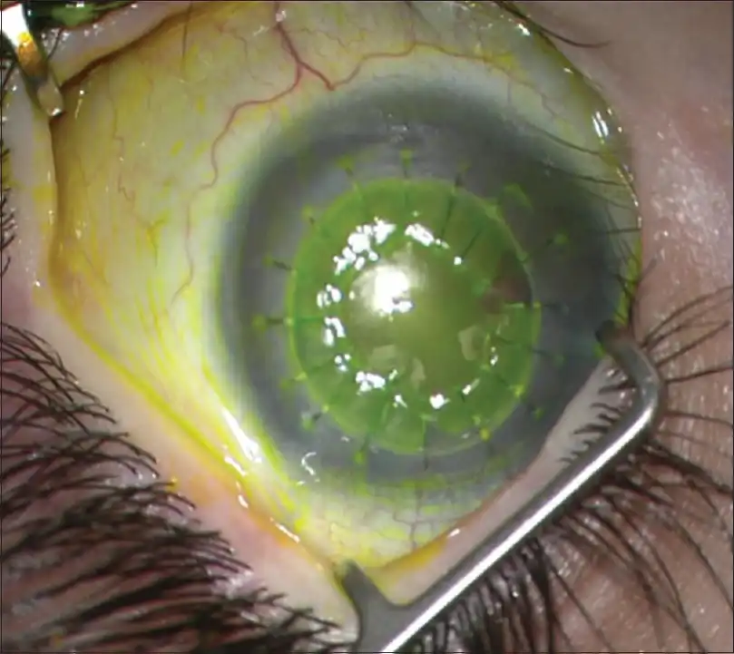
Figure 1
Left eye, positive fluorescein staining in a non-epithelialized corneal graft. 1 month post PKP
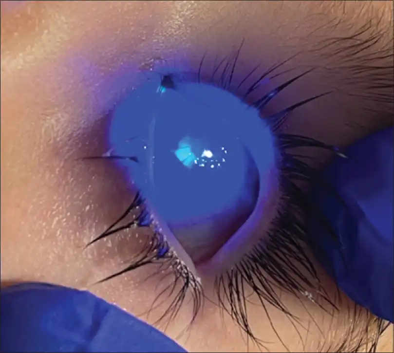
Figure 2
Left eye, 2 weeks after BioD placement. Graft more than 80% epithelialized with a small epithelial defect in the inferotemporal graft
Discussion
We present a case of corneal transplant, where the graft failed to epithelialize during a concomitant infection of confirmed COVID-19 by positive polymerase chain reaction (PCR) for SARS CoV-2. Prior to surgery, the patient did not have signs of stem cell deficiency, medication toxicity, and vitamin deficiencies.
SARS-CoV-2 infection requires ACE2 receptors to penetrate host cells.[] Zhou et al.[] demonstrated the positivity of ACE2 receptor in the corneal epithelium, endothelium with predominance in the limbal area. Because SARS-CoV-2 induces a dysregulated cytokine release,[] we hypothesize that the virus caused limbal inflammation that resulted in a lack of epithelialization as described in our case. We understand that delayed epithelial healing can be seen in pediatric patients with congenital glaucoma undergoing a corneal transplant. Because the COVID-19 infection correlated with the immediate postoperative period and the epithelium in the fellow eye showed an unhealthy epithelium after the infection, we think COVID-19 should be considered as a possible cause of the delayed epithelial healing. While there have been some reports of acute graft rejection,[] this is the first report of failed epithelization in a graft in a patient with COVID-19 infection.
Financial support and sponsorship
This publication was partially supported by an unrestricted grant from the Research to Prevent Blindness (RPB) Foundation to the Flaum Eye Institute at the University of Rochester.
Conflicts of interest
There are no conflicts of interest.
References
1
Sen M, Honavar SG, Sharma N, Sachdev MS COVID-19 and eye: A review of ophthalmic manifestations of COVID-19 Indian J Ophthalmol 2021 69 488 5092
Sindhuja K, Lomi N, Asif MI, Tandon R Clinical profile and prevalence of conjunctivitis in mild COVID-19 patients in a tertiary care COVID-19 hospital: A retrospective cross-sectional study Indian J Ophthalmol 2020 68 1546 503
Meduri A, Oliverio GW, Mancuso G, Giuffrida A, Guarneri C, Venanzi Rullo E, et al. Ocular surface manifestation of COVID-19 and tear film analysis Sci Rep 2020 10 1 7 doi: 10.1038/s41598-020-77194-94
Loden JC, Price FW Jr Price graft-over-host technique to manage positive pressure during penetrating keratoplasty J Cataract Refract Surg 1998 24 736 85
Zhou L, Xu Z, Castiglione GM, Soiberman US, Eberhart CG, Duh EJ ACE2 and TMPRSS2 are expressed on the human ocular surface, suggesting susceptibility to SARS-CoV-2 infection bioRxiv 2020 doi: 10.1101/2020.05.09.0861656
De Freitas Santoro D, de Sousa LB, Câmara NO, de Freitas D, de Oliveira LA SARS-COV-2 and ocular surface: From physiology to pathology, a route to understand transmission and disease Front Physiol 2021 12 612319 doi: 10.3389/fphys.2021.6123197
García LF Immune response, inflammation, and the clinical spectrum of COVID-19 Front Immunol 2020 11 1441 doi: 10.3389/fimmu.2020.014418
Jin SX, Juthani VV Acute corneal endothelial graft rejection with coinciding COVID-19 infection Cornea 2021 40 123 49
Singh G, Mathur U Acute graft rejection in a COVID-19 patient: Co-incidence or causal association? Indian J Ophthalmol 2021 69 985 6