Legends for Illustration
Ca: calcium
Er,Cr:YSGG: erbium, chromium:yttrium–scandium–gallium-garnet
Er:YAG: erbium:yttrium–aluminum-garnet
ICP-AES: ion-coupled plasma atomic emission spectrometer
P: phosphorus
Introduction
Direct bonding of brackets has revolutionized the clinical practice of orthodontics. The most common method of enamel preparation is phosphoric acid etching. However, acid etching removes the fluoride-rich surface layer of the enamel, thereby causing demineralization, which makes them more permeable and prone to acid attacks., This has been suggested as one of the reasons for the high prevalence of white spot lesions after orthodontic treatment with fixed appliances.
Alternative conditioning methods have been tried with poly(acrylic acid) or pre-treatment of the enamel surface with a sandblast of aluminum oxide, but these methods failed to achieve adequate bond strength to resist intraoral forces.,
Similarly different types of lasers such as CO2, erbium: yttrium–aluminum-garnet (Er:YAG), neodymium: yttrium–aluminum-garnet (Nd:YAG), and erbium, chromium: yttrium–scandium–gallium-garnet (Er,Cr:YSGG) have been used in orthodontics for enamel conditioning to bond brackets. However, the major disadvantage of these lasers was thermal damage, rendering these lasers unsuitable for hard tissue treatments. The innovation of Er:YAG laser and Er,Cr:YSGG laser permit ablation without any thermal side effects.
Er,Cr:YSGG (2790 nm) has a high absorption coefficient of water in enamel since laser wavelengths operate in the region of the major absorption peak for water (2790 nm) and are thus suitable for hard-tissue ablation treatments. This has led researchers to explore its use in enamel conditioning.
Berk et al compared laser-irradiated enamel surfaces with different power outputs (0.5 W, 0.75 W, 1 W, 1.5 W, and 2 W with a constant frequency of 20 Hz) with conventional phosphoric acid etching. He found that low-powered laser irradiations (0.5 W, 0.75 W, and 1 W) were not capable of etching enamel surface, but he suggested that a dosimetry of 1.5- and 2-W laser irradiation may be an alternative to conventional acid etching.
It has been perceived that the ideal dosimetry for enamel conditioning with laser are predominantly 1.5 W/20 Hz, 2 W/20 Hz.- But up until now, no studies have been undertaken to evaluate the effect of frequency variations. Majority of the studies have focused on varying the power outputs, but frequency settings were kept constant at 20 Hz. When the frequency is increased, there is a possibility of increasing the number of contacts in the enamel, thereby increasing the surface area with decreased surface roughness.
In this context, the present study was conducted to evaluate the shear bond strength and demineralization resistance of enamel surface conditioned using Er,Cr:YSGG laser with a constant power output of 2 W but with variable frequencies of 15 Hz and 25 Hz, and then they were compared with conventional acid etching that was kept as a control.
Materials and Methods
The advantage of the Er,Cr:YSGG (BiolaseTM and WaterLase iPlusTM) laser used in this study is that the frequency settings are adjustable unlike the previous laser systems, where the frequency setting was fixed at 20 Hz.- Laser irradiation was carried out in non-contact mode at a distance of 5 to 7 mm with the equipment set at the configuration of 60% air and 30% water.
Sample Description
Sixty sound human premolars, with intact enamel surface, extracted for orthodontic reasons, were selected for the study. The teeth were cleared of soft tissue debris and blood and immediately stored in distilled water. Thirty teeth were randomly allocated to test 2 different parameters, viz. demineralization resistance and shear bond strength.
Sample Preparation for Testing Demineralization Resistance
Color-Coded Acrylic Blocks
Evaluation of Demineralization Resistance
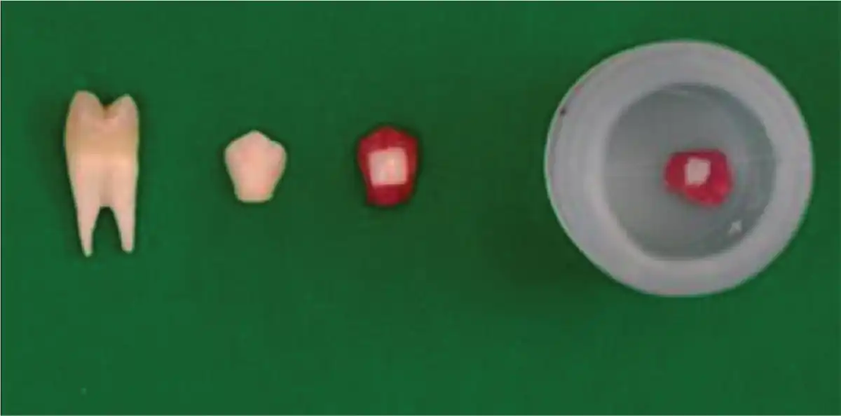
The crowns of 30 selected teeth were sectioned as presented in Figure 1. The complete enamel surface of the sectioned teeth was covered with nail varnish, except 5 mm × 5 mm window on the buccal surface and then assigned into 3 groups of 10 each.
Group A: Enamel etched with 37% phosphoric acid for 30 sec, and then rinsed and dried with oil-free water air spray for 15 sec and 10 sec each, respectively.
Group B: Enamel irradiated with Er,Cr:YSSG laser for 30 sec with power output of 2 W/15 Hz.
Group C: Enamel irradiated with the Er,Cr:YSSG laser with a power output of 2 W/25 Hz for 30 sec.
Demineralization solution was prepared by diluting nitric acid to 5% solution. Each prepared tooth was put into a separate container, containing 15 mL of demineralization solution and stored at room temperature for 24 h. Then, the solution was diluted 100-fold, and the dissolved calcium (Ca) and phosphorus (P) concentrations were measured with an ion-coupled plasma atomic emission spectrometer (ICP-AES. Acquired values were then subjected to statistical analysis.
Evaluation of Shear Bond Strength
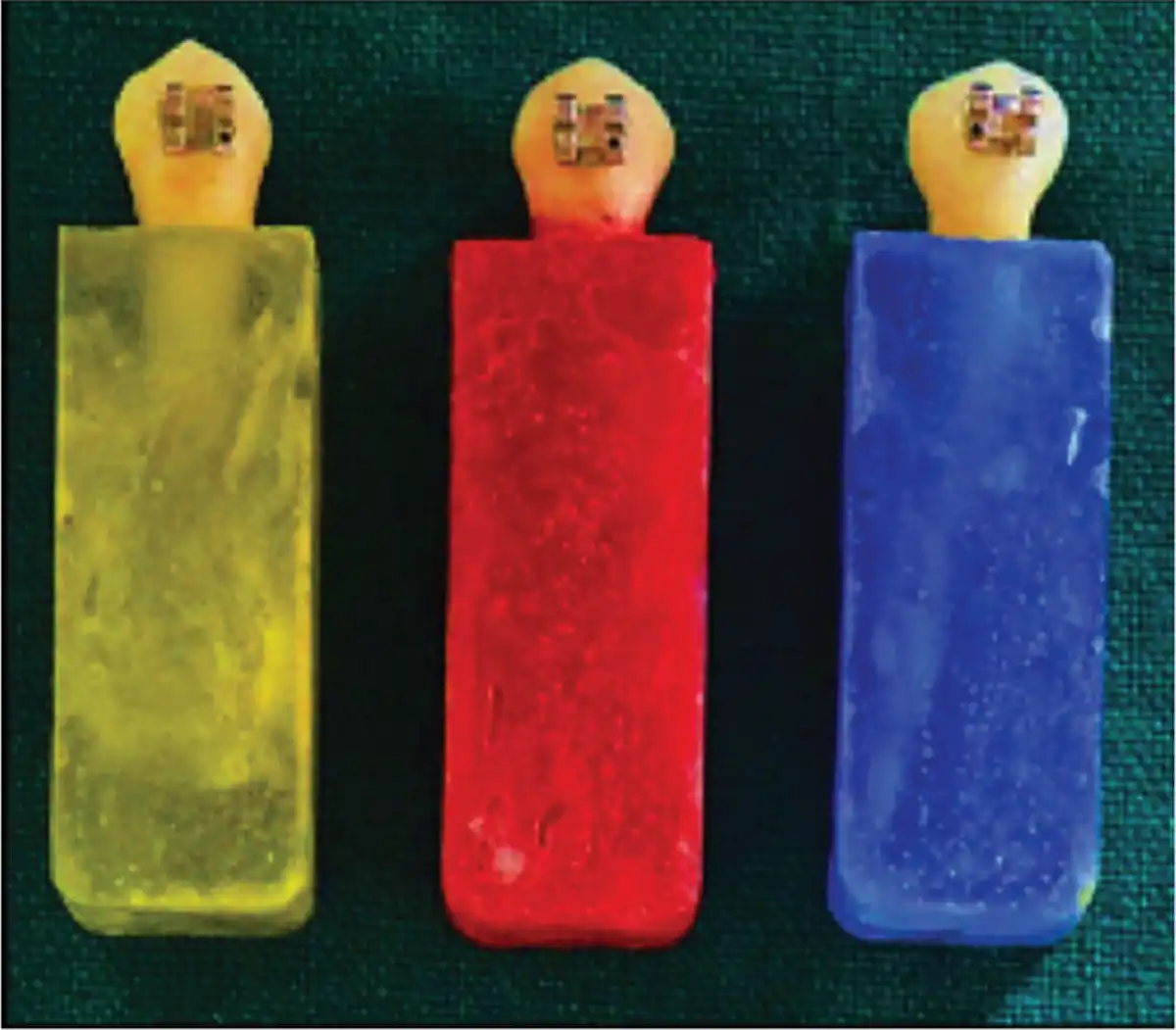
Thirty teeth were randomly allocated and embedded in color-coded acrylic blocks, which were divided into 3 groups of 10 teeth each for testing shear bond strength (Figure 2):
Group A (green blocks): Enamel etched with 37% phosphoric acid (3M, Dental products, St. Poul, USA) for 30 sec.
Group B (red blocks): Enamel irradiated with the Er,Cr:YSSG laser with a power output of 2 W, frequency of 15 Hz for 30 sec.
Group C (blue blocks): Enamel irradiated with the Er,Cr:YSSG laser with a power output of 2 W, frequency of 25 Hz for 30 sec.
After enamel conditioning, pre-adjusted edge-wise metal premolar brackets (Gemini 3M) were bonded onto the tooth surface with Transbond XT (3M Unitek, Monrovia, CA), and the samples were stored in distilled water at room temperature for 24 h for maintaining the hydration level. The samples were then tested for shear bond strength using a universal testing machine (INSTRON no. 3382) at cross-head speed of 0.5 mm/min force passing parallel to the buccal surface and the obtained data were subjected to statistical analysis.
After de-bonding, the enamel surface was observed under stereomicroscope (10× magnification, Olympus, SZX9, Olympus Corporation, Shinjuku-Ku, Japan), and the amount of remaining adhesive was evaluated according adhesive remnant index (ARI) developed by Artun and Bergland.
Results
The data collected were statistically analyzed using SPSS version 19.0 (IBM, Armonk, NY, released 2010).
Results of normality tests—Kolmogorov–Smirnov and Shapiro–Wilks—showed that all the variables followed normal distribution. To compare mean values between the 3 groups, one-way ANOVA was used, followed by Tukey’s HSD post hoc tests for pair-wise comparison. (If P-value was < .05, then it was considered to be statistically significant).
Evaluation of Demineralization Resistance
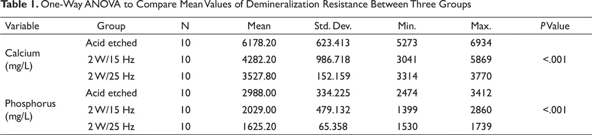
Highest dissolved mean Ca and P was found in the acid-etched group (Ca—6178.20 mg/L, P—2988 mg/L), followed by 2 W/15 Hz laser-etched group (Ca—4282.20 mg/L, P—2029 mg/L), and least with 2 W/25 Hz laser-etched group (Ca—3527.80 mg/L, P—1625 mg/L). The results were statistically significant (Table 1).
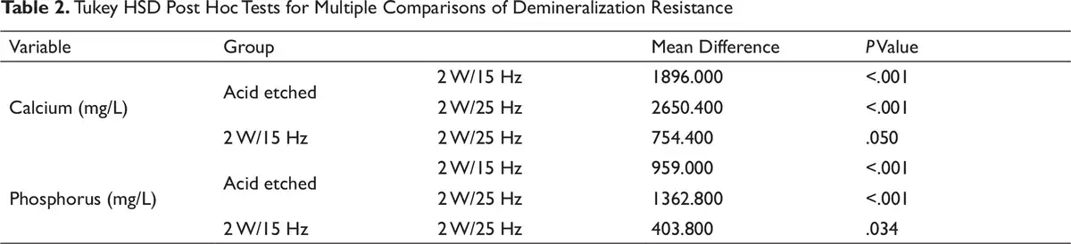
Tukey HSD post hoc tests revealed that there were statistically significant differences, when comparing the acid-etched group with 2 W/15 Hz and 2 W/25 Hz laser-etched group (P < .001). Whereas among laser-etched group, the difference was not statistically significant (P > .05). (Table 2).
Assessment of Shear Bond Strength
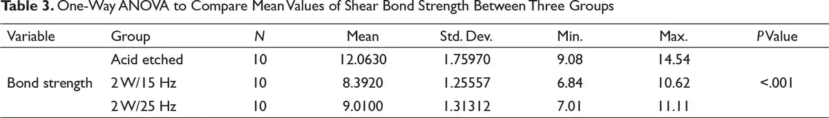
One-way ANOVA was performed to compare the mean values among 3 groups. There were statistically significant difference (P < .001) in bond strength between the groups, and the highest bond strength was found in the acid-etched group (12.06 ± 1.75 MPa), followed by 2 W/25 Hz (9.01 ± 1.3 MPa) and then 2 W/15 Hz laser-etched group (8.39 ± 1.25 MPa). (Table 3)
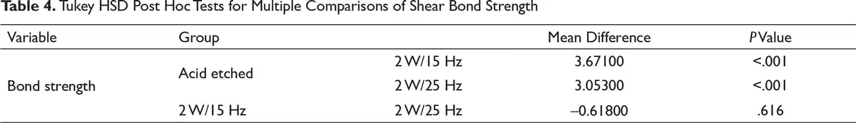
Tukey HSD post hoc tests were performed for multiple pair-wise comparisons. There were statistically significant differences while comparing acid-etched group with 2 W/15 Hz and 2 W/25 Hz laser-etched group (P < .001). No statistically significant differences were found between the laser-etched groups (P > .05). (Table 4).
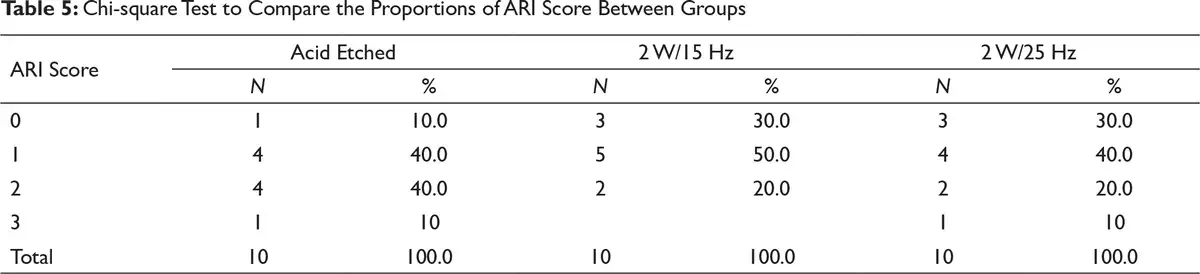
The ARI scores of the samples without saliva contamination are listed in Table 5. On acid-etched surfaces, 80% of the bond failure sites were within the adhesive. On 2 W/15 Hz laser-etched surfaces, 70% of the bond failure sites were within the adhesive. And on 2 W/25 Hz laser-etched surfaces, 60% of the bond failure sites were within the adhesive. However, the results were statistically not significant (P > .05).
Discussion
The demineralization resistance and the shear bond strength between acid-etched and laser-etched enamel surface was evaluated using ICP-AES and Universal Testing Machine, respectively.
On comparing the 3 groups, there was statistically significant difference in the concentration of dissolved Ca and P, with the highest dissolution in the acid-etched group, followed by 2 W/15 Hz laser-etched group, and least with 2 W/25 Hz laser-etched group (Table 1).
This findings are in agreement with the studies of Hossain et al and Kim et al, which revealed Er:YAG laser-treated enamels were more acid resistant due to improved crystalline structure and blocking effect of organic matrix when compared with acid-etched enamel.
The mechanisms underlying the demineralization resistance of laser can be due to:
Reduction in the carbonate content and modification of the organic matrix. In lased enamel, the inter-prismatic and intra-prismatic spaces that act as ion diffusion channels during the demineralization process were blocked by the decomposed organic materials, making the enamel less vulnerable to mineral loss., -
Formation of micro-spaces and micro-fissures in lased enamel. These spaces are believed to trap the Ca, P, and fluoride ions released from the tooth during the demineralization process.,
When comparing the shear bond strength, highest bond strength was found in the acid-etched group, followed by the 2 W/25 Hz and 2 W/15 Hz laser-etched group. The differences were statistically significant.
Comparatively lower bond strength with the laser than acid-etched group may be due to non-homogenous laser application with hand-sweeping motion, leaving untouched areas on the surface. 2 W/25 Hz laser-etched group showed increased bond strength compared to 2 W/15 Hz, and this may be attributed to the fact that there is a possibility of increasing the number of contacts in the enamel as you increase the frequency of the laser.
Maijer and Smith found a bond strength of 8 MPa to be adequate for orthodontic brackets; likewise, Reynolds proposed adequate bond forces range from 6 to 8 MPa. So, in this study, the shear bond strength values obtained from laser conditioning of enamel were clinically adequate.
Similar to our results, Ozer et al, Basaran et al, and Lee et al stated that laser etching yielded success rates as 37% phosphoric acid etching and did not find statistically significant differences in the laser- and acid-etched surfaces.
Nevertheless, the results of our study were in contrary to Usumez et al who reported that enamel conditioning with Er,Cr:YSGG laser was not a suitable method for orthodontic bonding .This could be due to the low laser power settings used in their study (1 W/20 Hz) that was not sufficient to create favorable etching surface.
Hence, it can be inferred that laser may be preferred over acid etching, as laser etching, in spite of providing optimum bond strength, increases demineralization resistance, thereby preventing white spot lesions which are the most prevalent iatrogenic consequence of orthodontic therapy.
Although all experimental steps of this study were conducted in a judicious manner and strictly according to the protocol, in vitro studies had some limitations to simulate oral environment. However, the main advantage of in vitro testing of demineralization resistance was that it provided investigators with the capability of performing single-variable experiments under controlled conditions. Several factors may contribute bracket bonding failure in patients. These factors were difficult to reproduce in the laboratory, and this could be one of the limitations of this study.
Hence, future studies should perform clinical trials on larger sample sizes and evaluate the precise efficacy of laser systems in terms of enamel conditioning.
Conclusion
It is prudent to conclude that Er,Cr:YSGG laser conditioning of enamel surface can be preferred over the conventional phosphoric acid etching in orthodontic bonding. Among the 2-laser parameters, 2 W/25Hz can be favored over 2 W/15 Hz because 2 W/25 Hz laser-etched surface yielded increased bond strength and more demineralization resistance potential over 2 W/15 Hz.
Acknowledgment
I owe enormous debt of gratitude and sincerely express my thanks to Dr Premila Suganthan BDS, MSc (Laser Dentistry), Director, KP Institute of Laser Studies, for her expert advice and encouragement throughout our research and permitting us to utilize Er,Cr:YSGG laser equipment in her institute.
Piradhiba R.
https://orcid.org/0000-0003-4161-6939
- 1. Chimello-Sousa DT, de Souza AE, Chinelatti MA, Pécora JD, Palma-Dibb RG, Milori Corona SA. Influence of Er:YAG laser irradiation distance on the bond strength of a restorative system to enamel. J Dent. 2006;34:245–251.
- 2. Øgaard B, Morten F. The enamel surface and bonding in orthodontics. Semin Orthod. 2010;16:37–48.
- 3. Retief DH, Dreyer CJ, Gavron G. The direct bonding of orthodontic attachments to teeth by means of an epoxy resin adhesive. Am J Orthod. 1970;58:21–40.
- 4. Medhi S, Mano MC, Sorel O, Cathelineau G. Enamel micro-abrasion. Orthod Fr. 2009;80:179–192.
- 5. Canay S, Kocadereli I, Akca E. The effect of enamel air abrasion on the retention of bonded metallic orthodontic brackets. Am J Orthod Dentofacial Orthop. 2000;117(1):15–19.
- 6. Jones SP, Gledhill JR, Davies EH. The crystal growth technique—a laboratory evaluation of bond strengths. Eur J Orthod. 1999;21(1):89–93.
- 7. Wigdor HA, Walsh JT, Featherstone JDB, Visuri SR, Fried D, Waldvogel JL. Lasers in dentistry. Lasers Surg Med. 1995;16:103–108.
- 8. Diaci1 J, Gaspirc B. Review: comparison of Er:YAG and Er,Cr:YSGG lasers used in dentistry. J Lasers Health Acad. 2012;2012(1):1–13.
- 9. Berk N, Basaran G, Ozer T. Comparison of sandblasting, laser irradiation, and conventional acid etching for orthodontic bonding of molar tubes. Eur J Orthod. 2008;30:183–189.
- 10. Basaran G, Ozer T, Berk N, Hamamci O. Etching enamel for orthodontics with an erbium, chromium:yttrium-scandium-gallium-garnet laser system. Angle Orthod. 2007;77:117–124.
- 11. Usümez S, Orhan M, Usümez A. Laser etching of enamel for direct bonding with an Er,Cr:YSGG hydrokinetic laser system. Am J Orthod Dentofac Orthop. 2002;122:649–656.
- 12. Usumez A, Ademci E, Usumez S. Effect of enamel laser irradiation at different pulse settings on shear bond strength of orthodontic brackets. Angle Orthod. 2013;83(6):973–980.
- 13. Artun J, Bergland S. Clinical trials with crystal growth conditioning as an alternative to acid etch enamel pre-treatment. Am J Orthod. 1984;84:333–340.
- 14. Hossain M, Nakamura Y, Yamada Y, Kimura Y, Nakamura G, Matsumoto K. Ablation depths and morphological changes in human enamel and dentin after Er,Cr:YSGG laser irritation in human enamel and dentin: ablation and morphological study. J Clin Laser Med Surg. 1999(b);17:155–161.
- 15. Kim JH, Kwon OW, Kim HI, Kwon YH. Acid resistance of erbium-doped yttrium aluminum garnet laser-treated and phosphoric acid-etched enamels. Angle Orthod. 2006;76:1052–1056.
- 16. Liu Y, Hsu CY. Laser-induced compositional changes on enamel: A FT-Raman study. J Dent. 2007;35:226–230.
- 17. Apel C, Meister J, Schmitt N, Graber HG, Gutknecht N. Calcium solubility of dental enamel following sub-ablative Er:YAG and Er:YSGG laser irradiatio. in vitro. Lasers Surg Med. 2002;30:337–498.
- 18. Liu JF, Liu Y, Stephen HC. Optimal Er: YAG laser energy for preventing enamel demineralization. J Dent. 2006;34:62–66.
- 19. Stern RH, Sognnaes RF, Goodman F. Laser effect on in vitro enamel permeability and solubility. J Am Dent Assoc. 1966;73:838–843.
- 20. Cecchini RC, Zezell DM, de Oliveira E, de Freitas PM, Eduardo Cde P. Effect of Er: YAG laser on enamel acid resistance: morphological and atomic spectrometry analysis. Lasers Surg Med. 2005;37:366–372.
- 21. Maijer R, Smith DC. Variables influencing the bond strength of metal orthodontic bracket bases. Am J Orthod Dentofac Orthop. 1981;79:20–34.
- 22. Reynolds IR. A review of direct orthodontic bonding. Br J Orthod 1975;2:171–178.
- 23. Ozer T, Başaran G, Berk N. Laser etching of enamel for orthodontic bonding. Am J Orthod Dentofacial Orthop. 2008;134(2):193–197.
- 24. Lee BS, Hsieh TT, Lee YL . Bond strength of orthodontic bracket after acid-etched, Er:YAG laser-irradiated and combined treatment on enamel surface. Angle Orthod. 2003;73:565–570.
- 25. Bishara SE, Ostby AW. White spot lesions: formation, prevention, and treatment. Semin Orthod. 2008;14(3):174–182.