Amyloid-like fibrils of ribonuclease A with three-dimensional domain-swapped and native-like structure
- Sambashivan, Shilpa
- Liu, Yanshun
- Sawaya, Michael R.
- Gingery, Mari
- Eisenberg, David
1Howard Hughes Medical Institute, UCLA-DOE Institute for Genomics and Proteomics, Box 951570, UCLA, Los Angeles, California 90095-1570, USA.
doi:10.1038/nature03916
†Present address: Division of Chemistry and Chemical Engineering, Caltech, Pasadena, California 91125, USA.
Received 18 January; accepted 16 June 2005.
Supplementary Information is linked to the online version of the paper at www.nature.com/nature.
Author Information The structure of Q10-H119A RNase A has been deposited in the Protein Data Bank with accession code 2APQ. The model of Fig. 4 has been deposited in the Protein Data Bank with accession code 2APU. Reprints and permissions information is available at npg.nature.com/reprintsandpermissions. The authors declare no competing financial interests. Correspondence and requests for materials should be addressed to D.E. ([email protected]).
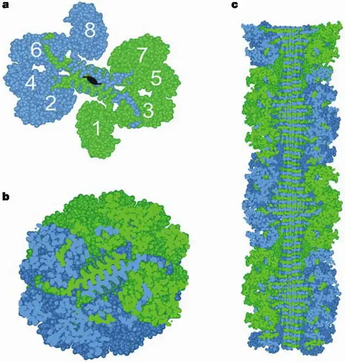
Figure 4
Domain-swapped zipper-spine model for the RNase A protofibril. a, The model is a 'runaway' domain swap between the RNase A monomers, with swaps occurring within one half protofibril but not between half protofibrils. Monomers 1-4 compose half the protofibrillar unit and are coloured as in Fig. 1c to emphasize domain swapping. The C-terminal β-strand of monomer 1 swaps into monomer 2, monomer 2 swaps into monomer 3, and monomer 3 swaps into 4, rising along the axis of the fibril. Q10 segments from these monomers form one antiparallel β-sheet in the spine. Monomers 5-8 form the other β-sheet, related to monomers 1-4 by a 21 axis along the fibril. Eight RNase A monomers comprise the asymmetric unit of the fibril. A similar model can be built from domain-swapped dimers; the currently available data do not favour one of these models over the other. b, The protofibril cross-section reveals the steric zipper, the interdigitation of Gln side chains in the spine of the fibril, modelled on the structure of GNNQQNY. c, A cut-away view perpendicular to the fibril axis reveals the stacking of hydrogen-bonded Q10 β-strands (4.88 Å apart) in the spine. The spine is largely shielded from solvent by the tight packing of globular domains around the periphery. The fibril model ranges from 100 to 140 Å in diameter, which agrees with the diameter of the fibrils obtained from electron microscopy images (Fig. 2, column 2).
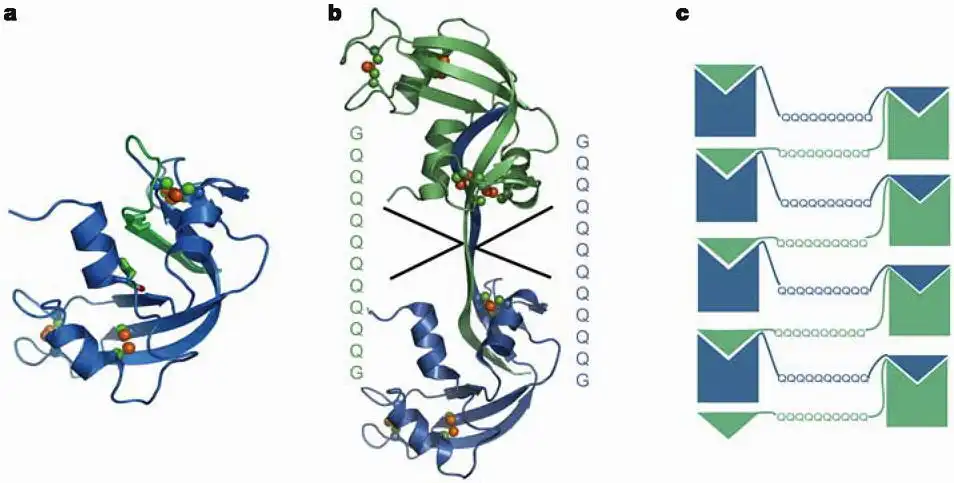
Figure 1
RNase A monomer and C-terminal domain-swapped dimer and the 3D domain-swapped zipper-spine model. a, The RNase A monomer is stabilized by four disulphide bonds Cys 26-Cys 84, Cys 40-Cys 95, Cys 58-Cys 110 and Cys 65-Cys 72, hindering conformational changes. His 12 in the core of the protein and His 119 on the β-strand that is swapped (shown by sticks) are active-site residues that we mutate to test for activity by complementation. b, The C-terminal domain-swapped dimer is formed by exchanging the C-terminal β-strands between two monomers. The hinge loop (residues 112-115) has been expanded by inserting the sequence -GQ10G-. c, Diagram of amyloid-like fibril formation in RNase A with Q10 expansion, leading to a runaway domain swap. The Q10-H12A mutants are shown in blue and the Q10-H119A mutants in green. Domain swapping between two mutants complements active sites.
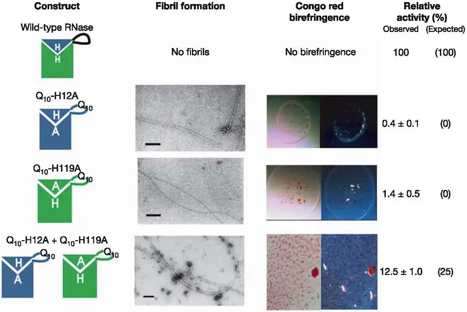
Figure 2
Properties of the RNase A amyloid-like fibrils. The hinge-loop region of wild-type RNase A that connects the C-terminal β-strand (triangle in the diagrams in column 1) to the protein core is expanded with the -GQ10G- motif to generate amyloid-forming RNase A mutants. Two inactive RNase A mutants are formed by replacing His 12 or His 119 with Ala. Wild-type RNase A does not form fibrils and has a fully functional active site (row 1, columns 2 and 4). The Q10-H12A, Q10-H119A and Q10-H12A + Q10-H119A constructs all form amyloid-like fibrils (column 2) and bind Congo red with the characteristic apple-green birefringence (column 3). The Q10-H12A + Q10-H119A fibrils (row 4, column 4) have significantly higher activity than fibrils of Q10-H12A (row 2, column 4) and Q10-H119A (row 3, column 4) alone. This is a result of complementation of active sites by domain swapping. The expected activity of each of the constructs is given in parentheses. Scale bar (column 2), 200 nm.
Amyloid or amyloid-like fibrils are elongated, insoluble protein aggregates, formedin vivoin association with neurodegenerative diseases orin vitrofrom soluble native proteins, respectively. The underlying structure of the fibrillar or 'cross-β' state has presented long-standing, fundamental puzzles of protein structure. These include whether fibril-forming proteins have two structurally distinct stable states, native and fibrillar, and whether all or only part of the native protein refolds as it converts to the fibrillar state. Here we show that a designed amyloid-like fibril of the well-characterized enzyme RNase A contains native-like molecules capable of enzymatic activity. In addition, these functional molecular units are formed from a core RNase A domain and a swapped complementary domain. These findings are consistent with the zipper-spine modelin which a cross-β spine is decorated with three-dimensional domain-swapped functional units, retaining native-like structure.